
A single visceral-fat number can mislead: the visceral adipose tissue normal range is often described as about 10% of total body fat, but interpretation depends on how it’s measured. For women, a waist measurement of 35 inches or more indicates increased risk of health problems related to visceral fat, according to the National Heart, Lung, and Blood Institute. Some body-composition monitors typically classify ratings from 1 to 12 as healthy and 13 or higher as high.
It’s easy to focus on what you see in the mirror. But visceral fat is stored deep in the abdomen, around internal organs, so appearance alone can’t show how much is present or what it means for your health.
This guide explains what visceral fat is, how to interpret common ranges and clinical thresholds, and why measurement methods can produce different results. You’ll also learn practical ways to reduce visceral fat, from consistent exercise and nutrition habits to tracking changes with objective body-composition data.
Key Takeaways
• Learn why waist measurements and device ratings are useful indicators, but shouldn’t be treated as interchangeable measures.
• Understand how visceral fat differs from fat stored just beneath the skin and why its location matters.
• See how clinical thresholds describe risk, not a complete picture of an individual’s health.
• Identify practical habits, including regular activity and dietary adjustments, that can support visceral-fat reduction.
• Explore how a DEXA scan can add objective body-composition data when you want a clearer baseline to track.
What is a healthy visceral fat range for a woman?
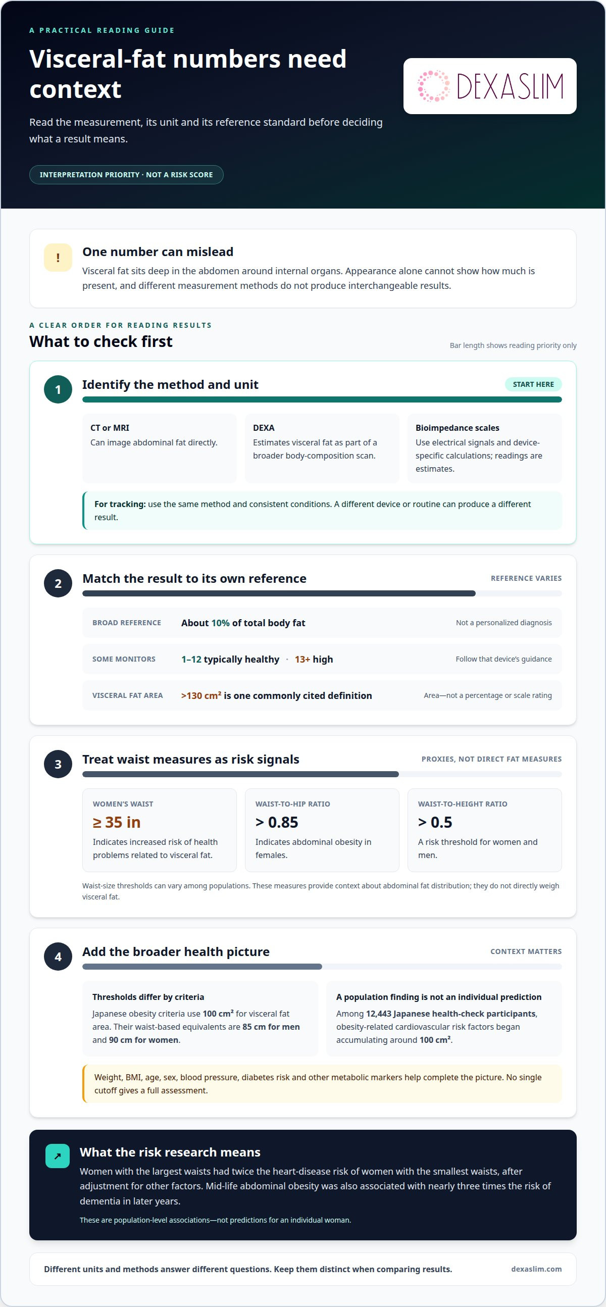
There isn’t one universal visceral-fat number that defines a healthy range for every woman. The result depends on the measurement method, body size, age, and the reference standard used. A waist measurement adds a practical risk signal: according to the National Heart, Lung, and Blood Institute, 35 inches or more in females indicates a risk of health problems related to visceral fat.
Some body-composition guidance describes visceral fat as about 10% of total body fat. This is a broad reference, not a personalized diagnosis. Device ratings also vary: some monitors describe scores from 1 to 12 as typically healthy, while scores of 13 or higher are considered high. Read a score alongside its device’s guidance, rather than treating every monitor’s scale as identical.
Waist measurements can help interpret risk. A waist-to-hip ratio above 0.85 indicates abdominal obesity in females, and a waist-to-height ratio above 0.5 is a risk threshold for women and men. Waist-size thresholds can vary among populations and are only one part of assessing health risk. These measures don’t directly weigh visceral fat. They offer context for how fat is stored around the abdomen, including the distinction between deeper visceral fat and subcutaneous fat beneath the skin.
Why does excess visceral fat matter? It is associated with cardiovascular and metabolic health risks. Women with the largest waists had twice the heart-disease risk of women with the smallest waists, after adjustment for other factors. Mid-life abdominal obesity was also associated with nearly three times the risk of dementia in later years. These are population-level associations, not predictions for an individual woman. Blood pressure, diabetes risk, family history, and other health measures help build a fuller picture.
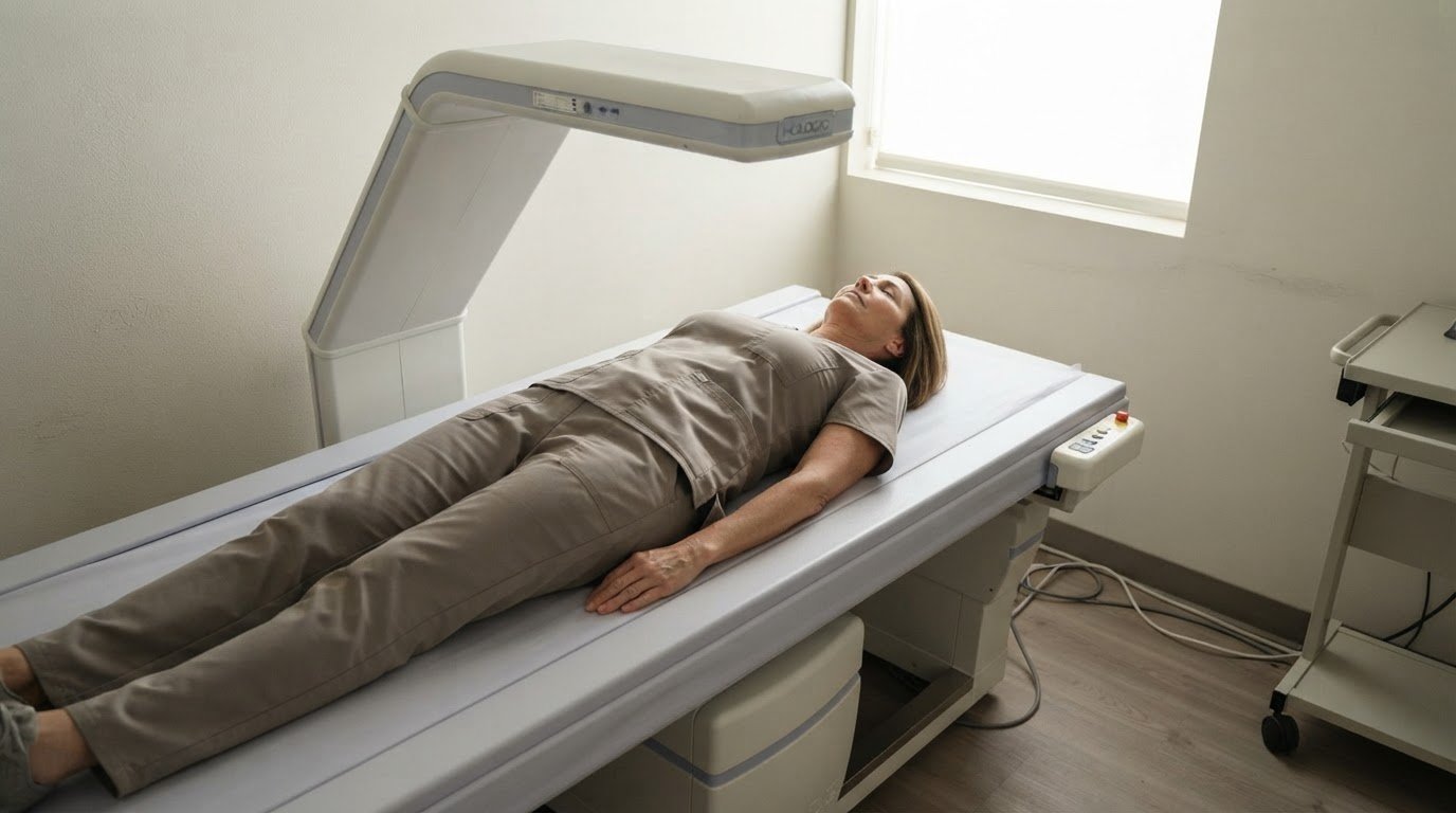
CT and MRI can image abdominal fat directly, while DEXA estimates visceral fat as part of a broader body-composition scan. Bioimpedance scales use electrical signals and device-specific calculations. Results aren’t interchangeable, so track changes using the same method and consistent conditions.
Consumer body-fat scales can help track a trend, but their visceral-fat readings are estimates, not direct images or clinical diagnoses. A different device or measurement routine can produce a different score.
Discuss a result with a healthcare professional if your waist measurement crosses a listed threshold, a monitor gives a high rating, or the result concerns you in light of blood pressure or diabetes risks. Bring the measurement method and result so they can be interpreted alongside your broader health profile.
Clinical visceral fat area threshold
Clinical visceral fat area thresholds describe abdominal fat measured inside the abdomen, rather than total body weight. One commonly cited definition classifies visceral obesity as visceral fat area exceeding 130 cm². This is an area measurement, not a percentage or a scale rating. Keep the unit attached: a result in cm² cannot be compared directly with pounds of fat or a monitor’s score.
Japanese obesity criteria use a different cutoff: 100 cm² defines visceral fat obesity. Those criteria use waist circumference as a practical equivalent, set at 85 cm for men and 90 cm for women. The waist measurement is a proxy, not a direct image of internal fat. It helps identify abdominal risk when area imaging isn’t the measure being used.
Why do the thresholds differ? They reflect different criteria and purposes, so one cutoff shouldn’t be treated as a universal boundary. A study of 12,443 Japanese health-check participants found obesity-related cardiovascular risk factors beginning to accumulate around 100 cm² of visceral fat area, irrespective of sex, age, or BMI. That finding describes a pattern across participants. It doesn’t mean every person at that measurement has the same metabolic risk.
Interpret the number in context. Weight and BMI describe overall body size, while visceral fat area focuses on fat within the abdominal cavity. Someone classified as overweight by BMI can have a different visceral fat level from someone with the same BMI. Age, sex, and individual metabolic markers also affect clinical interpretation. Insulin regulation, blood pressure, and other health indicators add context; the area figure alone cannot explain their causes.
Treat a threshold as a signal for assessment, not a diagnosis or guaranteed outcome. First, identify the method and unit on the report. Then distinguish a directly measured area from a waist-based estimate or device-generated rating. Finally, review the result alongside other available health information. This helps prevent a cm² cutoff from being mistaken for a complete assessment of abdominal obesity.
For a broader metabolic baseline, a Resting Metabolic Rate test measures resting energy expenditure; it doesn’t measure visceral fat area. That distinction matters when connecting body-composition findings with energy needs. Pairing the right metric with the right question keeps weight-management decisions grounded in what each measurement actually evaluates.
Visceral adipose tissue definition
Visceral adipose tissue, or VAT, is fat stored deep inside the abdomen, around organs such as the liver, pancreas, and intestines. It differs from subcutaneous fat, which sits beneath the skin and can be pinched around the waist or thighs. Both are body fat, but their location affects how they’re measured and interpreted.
VAT consists of fat cells, called adipocytes, within internal abdominal compartments. It isn’t the same as the visible softness around the stomach. For example, two people with similar waist circumferences may carry different amounts of visceral and subcutaneous fat. Appearance alone can’t distinguish these tissues or precisely measure visceral fat.
Visceral tissue has a biological role: it stores energy and participates in signaling processes involving hormones and other molecules. Excess accumulation is associated with metabolic and cardiovascular health risks, including diabetes risk. That association doesn’t make VAT a diagnosis of disease. It makes the amount and distribution of body fat relevant context for a broader medical assessment.
Think of body composition as a map, not a single weight reading. Body weight combines fat, muscle, bone, and other tissues. Waist circumference offers a simple clue about abdominal fat distribution, while imaging and device-based methods estimate VAT more directly. Each method answers a different question, so a waist measurement shouldn’t be mistaken for a direct VAT measurement.
Three distinctions help clarify reports:
Location
VAT surrounds organs inside the abdomen; subcutaneous fat lies beneath the skin.
Measure
A waist circumference records abdominal size, while imaging estimates internal fat quantity or area.
Meaning
A VAT result describes one part of body composition, not overall health or disease status.
Lifestyle can influence fat storage over time, but an individual result reflects more than one habit. Age, hormones, genetics, and overall body composition can affect fat distribution. A report is most useful when its method is clear and results are interpreted consistently. Comparing results from different methods can create confusion because each uses its own technique and scale.
Use the definition to guide your next decision: identify whether a result describes waist circumference, visceral-fat area, mass, or a device rating. Then consider what that measure can and can’t show. This keeps a single number in proportion and helps connect body-composition information with relevant metabolic markers and health priorities.
Ways to reduce visceral fat
Reducing visceral fat takes consistent changes to activity and food intake, not a single targeted exercise. Although abdominal fat surrounds organs, exercises can’t selectively remove fat from one area. Build a repeatable routine that supports overall fat loss and preserves muscle. Track waist circumference and body composition over time rather than judging progress by appearance alone.
A practical starting point is at least 30 minutes of exercise a day, five days a week. Choose cardio, such as brisk walking, or strength training that challenges major muscle groups. These approaches support an active routine and help make movement sustainable. Regular walking can also complement more structured workouts without requiring specialized equipment.
For a more structured plan, combine different training styles. Two HIIT workouts per week paired with daily walking are one suggested approach. Resistance training 2-4 days per week can help maintain or build muscle during weight loss. HIIT programs lasting 8-12 weeks can significantly reduce visceral fat. If considering high-intensity intervals, use a physiotherapist’s or sports coach’s supervision after a cardiac risk assessment.
Food choices matter alongside exercise. The Mediterranean diet and DASH diet are examples of eating approaches that can support a health-focused routine. A moderate calorie deficit of approximately 15-25 percent is another suggested target. Protein guidance is 0.7-1 gram per pound of goal body weight, or 1.6-2.2 grams per kilogram. These figures give structure, but food preferences and daily needs still shape a workable plan.
Recovery habits are part of the plan, too. Prioritize enough quality sleep and limit alcohol intake. These steps complement nutrition and training; they don’t replace them. Choose changes you can sustain through ordinary workdays, meals, and weekends instead of relying on short bursts of restriction or activity.
Measure progress consistently. Waist circumference can show whether abdominal size is changing, while body-composition measurements offer a different view of fat and lean tissue. Keep the method and routine consistent, then assess the trend rather than reacting to one reading. Consistent activity and managing food intake may lead to visible fat loss in two to three months.
If your plan includes a calorie deficit, protein targets, or intense exercise, align those choices with your health needs. A clinical assessment can add context when medical conditions, cardiovascular concerns, or medications affect exercise and nutrition decisions. Aim for steady progress by combining movement, food choices, sleep, and repeatable tracking. No single tactic is a complete solution for excess visceral fat.
What is visceral adipose tissue (VAT) and how to measure it?
Visceral adipose tissue, or VAT, is fat located among organs inside the abdominal cavity. A visceral adipose tissue normal range can’t be interpreted without knowing how the result was measured. Reports may describe VAT as mass, area, or a device rating. These outputs use different units and answer different questions. Identify the method and unit first, then determine whether the result comes from imaging or an estimate.
CT and MRI produce images that distinguish internal abdominal fat from nearby tissues. DEXA provides a broader body-composition scan and estimates visceral fat mass. Bioimpedance scales use electrical signals and their own calculation models to produce a rating. These methods aren’t interchangeable: a DEXA mass in pounds can’t be directly compared with an area in cm² or a scale score.
Age-specific reference values show why context matters. US DXA reference data report median VAT mass of 0.04 kg for women aged 18-19, 0.06 kg for women aged 20-24, 0.55 kg for women aged 25-49, and 1.21 kg for women aged 50 and older. For men, the medians are 0.18 kg, 0.25 kg, 0.71 kg, and 1.62 kg across those same age groups. These are reference medians, not individual targets. The visceral adipose tissue normal range therefore depends partly on the comparison group and measurement type.
A waist measurement is a practical proxy, not a scan. For consistent tracking, measure at the same location and under similar conditions each time. One recommendation is to record circumference to the nearest one-eighth inch or 0.5 cm. Tape placement can affect comparisons, so repeatable technique matters more than false precision.
Use this sequence to read a report:
Identify the method
CT, MRI, DEXA, bioimpedance, or tape measurement.
Check the unit
mass, area, circumference, or device rating.
Compare like with like
use the same method for follow-up trends.
Read the reference
age and sex can shape the comparison group.
Consumer scales can help track a device-specific trend, but a rating isn’t a direct image of internal fat. A sudden shift between devices may reflect different algorithms rather than a change in body composition. Keep the device and routine consistent, then interpret the result alongside other health information.
For an objective body-composition baseline, book a DEXA scan to assess fat distribution and track changes with a consistent method.
Dexaslim
Dexaslim helps turn body-composition questions into measurable health and performance data. A 1 Session Dexa Scan can provide a body-composition baseline, while VO2 Max Performance Testing evaluates aerobic endurance and cardiovascular efficiency. A Resting Metabolic Rate (RMR) Test measures resting energy expenditure, and a Longevity Panel adds broader health data.
Consider objective measurement if changes in waist size or a device rating leave you unsure how to track progress. Each assessment answers a different question, so choose based on the information you want to understand. Dexaslim offers DEXA body-composition scans to help you assess fat and lean tissue. Discuss which assessment best fits your goals.
Turn your measurements into a clear next step
Your visceral adipose tissue normal range is a reference point, not a complete health strategy. Use objective measurements to establish a baseline, then focus on consistent habits and meaningful progress over time. Choose information that matches your goals rather than collecting numbers without a purpose.
Dexaslim offers a 1 Session Dexa Scan for body-composition insight, VO2 Max Performance Testing to evaluate aerobic performance, and a Resting Metabolic Rate (RMR) Test to assess resting energy expenditure. Each provides a different perspective, helping you connect body composition with performance and metabolic data.
If you’re ready to make your next step data-informed, contact Dexaslim to discuss your options. Start with a clear question, choose measurements that address it, and use the results to guide your plan. Progress begins with a baseline you can understand and act on.
Frequently Asked Questions
What Is Visceral Fat?
Visceral fat is located within the abdomen, around internal organs. The key distinction is location: subcutaneous fat lies beneath the skin, while visceral fat sits deeper. A waist measurement can suggest abdominal fat distribution, but it can’t show exactly how much fat surrounds the organs. Imaging and body-composition methods estimate this internal fat in different ways, so the method matters when interpreting results.
Does visceral fat contribute significantly to body weight?
Visceral fat contributes to body weight, but it is only one component of total mass. Listed DEXA averages are approximately 1 pound for women and 2 pounds for men, based on NHANES 2017-2018 data. These averages don’t determine an individual’s result. A person’s scale weight also includes subcutaneous fat, muscle, bone, and other tissues, so weight alone can’t isolate visceral fat.
What are some conditions and disorders that affect visceral fat?
Obesity and metabolic health are relevant when assessing visceral fat. A BMI of 30 or more may indicate overweight and a higher level of visceral fat, but BMI doesn’t measure abdominal fat directly. Excess visceral fat is associated with cardiovascular and metabolic health risks. A result should therefore be considered alongside other health information, rather than used alone to identify a disorder or explain its cause.
What are the differences between visceral fat and subcutaneous fat?
Visceral fat lies deeper in the abdomen, while subcutaneous fat sits directly beneath the skin. For example, the soft tissue you can pinch around your waist is generally subcutaneous; it doesn’t reveal how much fat surrounds internal organs. Waist size and appearance can’t precisely distinguish the two. Imaging and body-composition methods estimate internal fat more directly.
What is the normal visceral fat area range in cm² for adults?
There isn’t one universal adult range in cm² that applies across all measurement systems. Japanese obesity criteria use 100 cm² as the threshold for visceral fat obesity, while another clinical definition describes visceral obesity as an area exceeding 130 cm². These are criteria, not interchangeable “normal ranges.” Check whether a report gives an area measurement, waist-based estimate, or another type of result.
What is a healthy visceral fat rating on a body composition scale?
On some body-composition monitors, ratings from 1 to 12 are typically described as healthy; a rating of 13 or higher is considered high and associated with increased health risks. These numbers are device ratings, not cm² or pounds. Use the scale’s own interpretation and compare readings from the same device over time. A score alone doesn’t diagnose a health condition.
What are normal VAT ranges by age and sex?
US DXA reference data report median VAT mass for women of 0.04 kg at ages 18-19, 0.06 kg at 20-24, 0.55 kg at 25-49, and 1.21 kg at 50 and older. For men, medians are 0.18 kg, 0.25 kg, 0.71 kg, and 1.62 kg across those same age groups. These are reference medians, not individual targets or diagnostic cutoffs.
What visceral fat level is associated with increased health risks?
Risk indicators depend on the measurement. Some monitors classify a visceral-fat rating of 13 or higher as high, while a visceral fat area of 100 cm² is the threshold used for obesity under Japanese criteria. Another definition places visceral obesity above 130 cm². These figures use different measures and criteria. Interpret the number with its method and other health markers, not as a standalone diagnosis.