
Your waistline can signal risk, but it cannot show exactly where abdominal fat is stored. The best way to measure visceral adipose tissue depends on whether you need a direct view of fat compartments or a practical estimate. MRI can image abdominal fat, while waist ratios and other body measurements offer indirect clues.
Visceral fat lies deep in the abdomen, around internal organs. It differs from subcutaneous fat, which sits just beneath the skin. A general body-fat estimate or BMI alone cannot tell you how much fat is visceral.
This article explains how waist circumference and waist ratios assess risk, what body-fat estimates can and cannot reveal, and how MRI compares with other methods. You will also learn what body measurements contribute and where a DEXA scan can fit into a broader body-composition assessment. Use these differences to choose a method that answers your question, whether you need a convenient screening measure or a more detailed view of abdominal fat.
Key Takeaways
• Use waist measurements and ratios to assess abdominal risk, but recognize that they don’t directly measure visceral fat.
• Understand why a total body-fat estimate can’t tell you precisely how much fat surrounds your organs.
• See how MRI offers a more direct view of abdominal fat compartments and how it compares with other methods.
• Learn what anthropometric measures can indicate and why BMI alone can’t identify visceral fat.
• Distinguish visceral fat around internal organs from subcutaneous fat beneath the skin to interpret measurements more clearly.
Waist-to-hip ratio
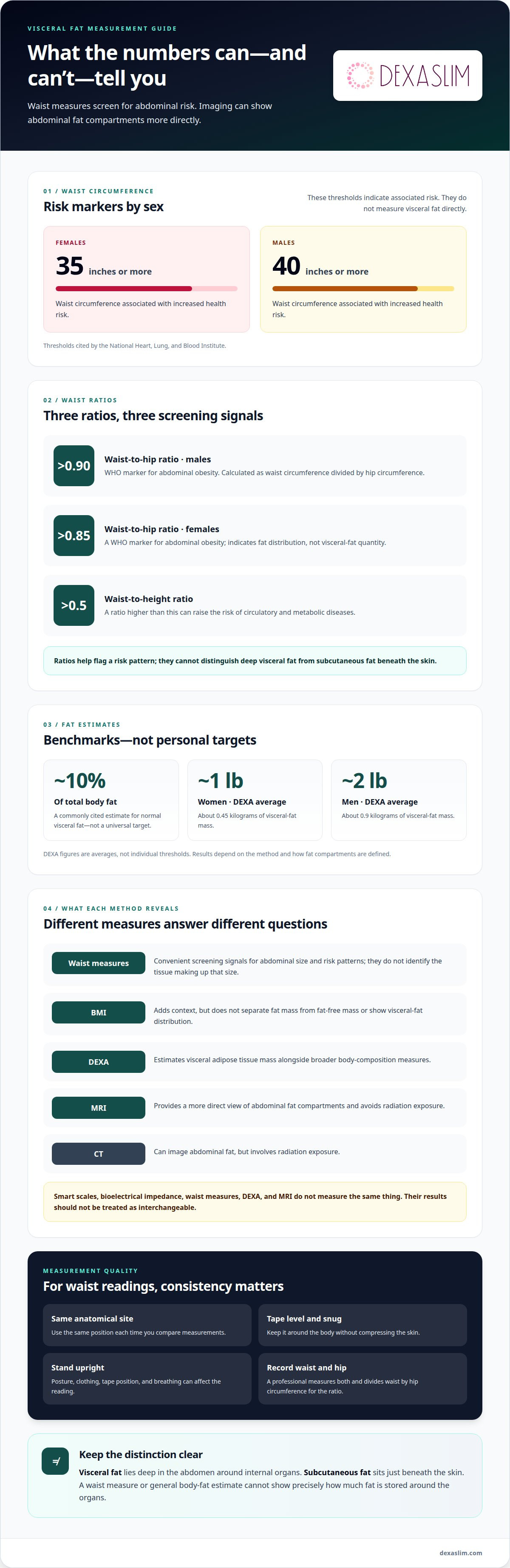
Waist-to-hip ratio compares waist circumference with hip circumference. A professional measures both and divides the waist measurement by the hip measurement. The result describes fat distribution, not the amount of visceral fat surrounding the organs. A ratio can flag an abdominal pattern associated with health risk, but it cannot distinguish deep visceral fat from subcutaneous fat beneath the skin. Treat it as one screening measure, not a complete body-composition assessment.
For females, a waist circumference of 35 inches or more indicates risk of health problems associated with visceral fat, according to the National Heart, Lung, and Blood Institute. For males, the corresponding risk threshold is 40 inches or more, according to the National Heart, Lung, and Blood Institute. The World Health Organization identifies a waist-to-hip ratio above 0.90 in males as abdominal obesity; a ratio above 0.85 in females is also used as a marker. These are risk thresholds, not direct measurements of visceral fat.
What visceral fat measurements or ranges are considered healthy by age and sex? The cited markers are organized by sex, not presented as age-specific healthy visceral-fat ranges. They indicate associated risk, rather than a precise visceral-fat quantity or a universal healthy range. A waist-to-height ratio higher than 0.5 can raise the risk of circulatory and metabolic diseases. Unlike waist-to-hip ratio, this measure compares waist circumference with height, making body proportions part of the interpretation.
For consistent results, a professional uses the same anatomical site and keeps the tape level around the body. You stand upright while the tape sits snugly without compressing the skin. The professional records waist and hip circumferences, then calculates the ratio. Clothing, posture, tape position, and breathing can affect the reading. Consistent positioning is important when comparing results over time, especially if weight or lifestyle changes may affect waist size.
BMI adds context, but it does not distinguish fat mass from fat-free mass. A muscular person and a person with more body fat can have a similar BMI, so BMI alone cannot show visceral-fat distribution. Waist circumference also has limits: it reflects abdominal size, not the tissue that makes up that size. CT involves radiation exposure, while MRI avoids radiation, making MRI useful for repeated imaging comparisons.
Use waist measurements as accessible screening signals, then interpret them alongside other body-composition data. Crossing a threshold can prompt a closer assessment of cardiovascular and metabolic health, but it does not diagnose the amount of visceral fat. The best way to measure visceral adipose tissue depends on your question: waist ratios track risk patterns, while imaging assesses abdominal fat compartments more directly. Choose the measure that fits the decision you need to make.
Estimating visceral fat as a share of total body fat
Total body fat combines fat stored throughout the body, but visceral fat is only one compartment. A commonly cited benchmark describes normal visceral fat as about 10% of total body fat. Treat that percentage as an estimate, not a universal target. Measurement methods define and calculate fat compartments differently, so a percentage can’t be interpreted apart from the method and its reported units.
DEXA estimates visceral adipose tissue mass alongside broader body-composition measures. Reported average DEXA visceral-fat mass is about 1 pound (0.45 kilograms) for women and about 2 pounds (0.9 kilograms) for men. These are averages, not individual thresholds. A scan result should be read as a method-specific estimate, rather than proof that a person’s visceral fat matches a fixed share of total body fat.
Total body-fat estimates have limits, too. They describe fat mass in relation to the rest of the body, but do not show where that fat is stored. BMI is less specific still because it does not separate fat mass from fat-free mass. For example, two people with similar body weight can have different body composition and abdominal fat distribution. This distinction matters when assessing obesity or metabolic risks associated with visceral fat and diabetes.
How much do CT, MRI, and DEXA visceral fat scans cost in the United States? Costs depend on scan type and the specific case; DEXA pricing also varies by the service provided. The cost of imaging can reflect the scan protocol and the analysis involved. Insurance coverage is governed by plan benefits and the clinical indication for the scan.
How reliable are professional DEXA scans compared with smart scales, bioelectrical impedance, and waist measurements? DEXA provides a scan-based estimate, while smart scales and bioelectrical impedance estimate composition indirectly, and waist measurements assess abdominal size. These methods don’t measure the same thing. A study comparing bioelectrical impedance and ultrasound estimates with MRI illustrates why their results shouldn’t be treated as interchangeable or as exact measures of visceral-fat mass.
How can you get a measurement, and how should you prepare? A professional selects the method based on whether the goal is a broad body-composition estimate or more focused imaging. The scan procedure and preparation follow the selected method’s protocol; the professional positions the person, acquires the measurement, and explains the resulting estimate. For metabolic context alongside body-composition data, a Resting Metabolic Rate (RMR) Test measures resting energy needs, but doesn’t measure visceral fat.
A commonly cited benchmark is visceral fat at about 10% of total body fat, but it is an estimate rather than a universal target. DEXA averages are about 1 pound (0.45 kilograms) for women and 2 pounds (0.9 kilograms) for men; these are averages, not individual thresholds, and no age-specific ranges are provided here.
MRI as a visceral fat comparator
MRI and CT estimate visceral fat by imaging abdominal tissue and separating fat compartments. A trained professional or image-analysis system identifies visceral adipose tissue inside the abdominal cavity, distinct from subcutaneous fat beneath the skin. The resulting area or volume depends on scan coverage, image quality, and the boundary used. Neither a waist circumference nor a scan duration in minutes describes those choices.
CT and MRI create images that can be analyzed to distinguish abdominal fat from surrounding structures. Accuracy depends on segmentation rules and scan coverage. Measurements across multiple slices represent compartment volume more fully than a single-slice assessment.
A single MRI slice can estimate abdominal adipose tissue at a standardized location. Studies have identified a level about 5-10 cm above L4-L5, around L3. On a transverse image, the inner boundary of the abdominal muscle wall helps define visceral adipose tissue. This consistent landmark supports comparisons, but a single slice remains a sample of the abdomen, not a complete volume measurement.
For fuller quantification, whole-body or regional multi-slice imaging captures fat across multiple sections. Scan ranges can extend from the top of the liver to the pubic symphysis, or from the top of the liver to the bottom of the pelvis. These approaches measure compartment volumes more fully than single-slice methods. Broader coverage can clarify how visceral and subcutaneous fat are distributed across the abdomen.
CT analysis also uses tissue-density thresholds, expressed in Hounsfield units. One described segmentation range is -195 to -45 Hounsfield units; another reported range is -190 to -30 Hounsfield units. Different thresholds can affect which pixels count as adipose tissue. A clinical result therefore depends not only on the image, but also on the segmentation protocol and the anatomical boundaries applied.
MRI avoids radiation exposure, making it suitable for multi-slice longitudinal studies. CT involves radiation exposure, so its use depends on the clinical question and the value of its imaging data. Neither modality alone predicts whether someone will develop a disease. Imaging quantifies fat compartments; clinicians interpret those measurements alongside other health information, rather than treating a scan result as a complete assessment.
Technique matters. An anatomical method for estimating the boundary between intra- and extraperitoneal fat showed errors of 3.8-49.4% compared with high-resolution MRI. That wide range demonstrates how boundary selection can shift estimates. For reliable comparisons over an interval, consistent scan levels, coverage, and segmentation rules are essential. A waist measure can show external circumference, but MRI or CT reveals internal compartments directly.
The process involves imaging abdominal tissue, then identifying visceral fat inside the abdominal cavity separately from subcutaneous fat. A professional or image-analysis system evaluates the images. The measured area or volume depends on scan coverage, image quality, and the boundaries used.
Anthropometric visceral fat assessment
Anthropometric models estimate visceral adipose tissue from body measurements without CT or DEXA scans. A professional records dimensions such as height, weight, and abdominal circumference, then applies a model to estimate visceral fat. The result is an estimate, not an image or direct measurement of fat around the organs. Its usefulness depends on the model and the accuracy of the measurements entered.
That distinction is important. A prediction may help identify an abdominal fat pattern, but it cannot show precisely where visceral fat ends and subcutaneous fat begins. Two people with similar body measurements may still have different fat distributions. The model turns measurable body dimensions into an estimate; it does not examine internal tissue. Use it as one part of an assessment, not as a substitute for imaging when direct compartment measurement is needed.
BMI illustrates both the value and the limits of anthropometry. A BMI of 30 or more may indicate overweight and could accompany a higher level of visceral fat. Yet BMI does not indicate visceral fat, which can be a chronic-disease risk factor independently of total fat mass. It also cannot distinguish fat from fat-free mass. A professional therefore interprets BMI with circumference and other relevant measures, rather than treating one number as a complete body-composition profile.
Consistent measurements affect the estimate. During an assessment, the practitioner uses a tape measure and a defined protocol to record each body dimension. Posture, tape placement, clothing, and tension can change a circumference reading, affecting the model’s result. For meaningful comparisons across visits, use the same measurement approach and recording conditions each time.
Anthropometric assessment works best as a practical screening tool. It can help track changes in abdominal measurements, even though it does not directly quantify visceral fat. For example, a stable BMI alongside a changing waist measurement can signal a shift in body shape that BMI alone misses. That pattern still does not identify the exact amount of visceral fat or establish a diagnosis.
Interpret the estimate in context. Consider the measurements, the model’s intended use, and the question you want answered. If your goal is to screen for a possible abdominal risk pattern, anthropometric data can be useful. If you want to quantify an internal fat compartment, imaging offers a more direct assessment. The best way to measure visceral adipose tissue depends on whether you need accessible screening or a more detailed tissue assessment.
Visceral adipose tissue and its location
Visceral adipose tissue, or VAT, lies deep inside the abdomen around internal organs. It differs from subcutaneous fat, which sits beneath the skin and can be pinched externally. A waist measurement reflects abdominal size, but cannot show which tissue contributes to that circumference. Imaging adds a view of internal fat compartments, showing where fat is distributed instead of relying only on body shape.
Location matters because visceral fat occupies the abdominal cavity, not simply the area around the waist. In image analysis, professionals distinguish VAT from fat outside the abdominal muscle wall. The inner boundary of that wall helps define the visceral compartment on a cross-sectional image. This anatomical landmark makes the result more specific than a tape measure, which records an external dimension without separating internal and subcutaneous fat.
VAT is one part of a broader body-composition picture. Total body-fat estimates combine fat across regions, while visceral-fat measurements focus on a particular abdominal compartment. A DEXA scan can estimate visceral-fat mass alongside other body-composition measures. MRI can show abdominal fat compartments across selected slices or broader scan coverage. These methods answer different questions, so choose the measurement that matches your goal: total composition, abdominal distribution, or change over time.
How often should visceral adipose tissue be measured to track changes? A DEXA scan every 3-4 months can assess changes in visceral fat, lean mass, and bone health. Waist circumference can be remeasured every 4-6 weeks to follow a simpler external trend. Keep the method consistent across assessments; switching from a circumference measure to imaging changes what the result represents.
Interpret location data alongside the factors that shape it. Body weight alone cannot reveal whether abdominal fat is visceral or subcutaneous. Exercise and lifestyle changes may alter body composition, but the measurement method determines which compartment a follow-up can track. A smaller waist, for example, indicates a change in abdominal circumference, while imaging can assess visceral-fat mass more directly. Neither result alone diagnoses chronic disease, cancer, or an inflammatory condition.
The clearest takeaway is simple: visceral fat is defined by where it sits, not by how the abdomen looks. A circumference is useful for tracking external change; imaging evaluates internal distribution. Scan coverage and analysis boundaries also influence the result, so consistent methods matter when comparing measurements across an interval. To establish a measurable body-composition baseline, book a DEXA scan.
Dexaslim
Dexaslim helps turn body-composition questions into measurable health data. If you want a professional assessment after comparing waist measures and imaging methods, a 1 Session Dexa Scan can provide body-composition information, including a visceral-fat estimate. Pair that data with other evaluations when your goals extend beyond fat distribution: VO2 Max Performance Testing assesses aerobic performance, while a Resting Metabolic Rate (RMR) Test measures resting energy needs. A Longevity Panel adds another avenue for exploring health markers.
Choose the assessment that matches the question you want answered. Contact Dexaslim to discuss your measurement goals and the relevant evaluation.
Make Your Next Measurement Count
Choosing the best way to measure visceral adipose tissue starts with defining the decision your data needs to support. A screening signal, a body-composition baseline, and a detailed view of abdominal fat are different goals. Match the method to your question, then use a consistent approach to track change. This keeps results meaningful and helps prevent a change in measurement method from appearing to be a change in your body.
Turn the information into a practical next step. Consider what you want to understand, what type of result would answer your question, and how the measurement fits your broader health goals. Clear goals make it easier to interpret data and use it to guide future decisions.
Explore your measurement options with Dexaslim and take the next step with clarity. A more informed baseline can help you move forward with purpose.
Frequently Asked Questions
What Is Visceral Fat?
Visceral fat is stored deep within the abdomen, around internal organs. It differs from fat beneath the skin, which is called subcutaneous fat. Its location defines it, not simply a larger waist or higher body weight. A waist measurement can show abdominal size, while imaging can distinguish internal fat compartments. This difference clarifies what each measurement can and cannot tell you.
What is the most accurate way to measure visceral adipose tissue?
MRI with multi-slice imaging provides a detailed assessment of visceral-fat compartment volume without radiation exposure. Results depend on scan coverage and how image analysis defines the boundary between visceral and surrounding fat. For meaningful follow-up, compare scans with consistent coverage and analysis criteria. A single-slice MRI can estimate abdominal fat at a standard level, but samples less tissue than multi-slice imaging.
Can a DEXA scan measure visceral adipose tissue accurately?
A scan-based estimate should be interpreted as an estimate rather than a definitive measure of visceral adipose tissue. Interpret the estimate as part of the scan’s body-composition analysis, not as an exact count of fat around the organs. For tracking, use the same measurement method over time. Differences between technologies may reflect how each method estimates fat, not just changes in your body.
How much do CT, MRI, and DEXA visceral fat scans cost in the United States?
Costs can vary by scan type and service.
What visceral fat measurements or ranges are considered healthy by age and sex?
The material gives no age-specific healthy ranges. Risk markers include waist circumferences of 35 inches or more for females and 40 inches or more for males, and waist-to-hip ratios above 0.85 for females or 0.90 for males. About 10% of total body fat is a commonly cited estimate, not a universal target.
What are the risks of CT radiation when measuring visceral fat?
CT involves radiation exposure. MRI avoids radiation exposure and is described as safer for multi-slice longitudinal studies.